Преподавание специальности на додипломном и
реклама
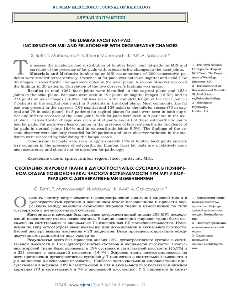
RUSSIAN ELECTRONIC JOURNAL OF RADIOLOGY СЛУЧАЙ ИЗ ПРАКТИКИ THE LUMBAR FACET FAT-PAD: INCIDENCE ON MRI AND RELATIONSHIP WITH DEGENERATIVE CHANGES T S. Butt1, T. Muthukumar1, S. Iftikhar Mahmood1 , K .Ali3, A. Saifuddin1,2 o assess the incidence and distribution of lumbar facet joint fat-pads on MRI and correlate of the presence of fat-pads with osteoarthritic changes in the facet joints. Materials and Methods: lumbar spine MRI examinations of 200 consecutive patients were studied retrospectively. Presence of fat pads was noted on sagittal and axial T1W SE images. Osteoarthritic changes were noted in the axial plane. A second observer recorded the findings in 20 patients. Correlation of the two observer’s findings was made. Results: in total 1261 facet joints were identified in the sagittal plane and 1534 joints in the axial plane. Fat-pads were seen in 194 joints on sagittal images (15,5%) and in 221 joints on axial images (14,4%). Fat was seen in the complete length of the facet joint in 7 patients in the sagittal plane and in 3 patients in the axial plane. Most commonly, the fat pad was present in the superior (109 sagittal and 134 axial) or the inferior recess (73 in sagittal and 76 in axial plane). In 5 patients (in sagittal plane) fat pads were seen in both superior and inferior recesses of the same joint. Such fat pads were seen in 8 patients in the axial plane. Osteoarthritic change was seen in 450 joints and 43 of these osteoarthritic joints had fat pads. Fat pads were less common in the presence of facet osteoarthritis (Incidence of fat pads in normal joints 16,4% and in osteoarthritic joints 9,5%). The findings of the second observer were similarly recorded for 20 patients and inter-observer variation in the statistics were recorded by calculating the kappa scores. Conclusion: fat pads were seen in approximately 15% of lumbar facet joints and are less common in the presence of osteoarthritis. Lumbar facet fat pads are a relatively common occurrence and should not be mistaken for pathology. 1 - The Royal National Orthopaedic Hospital NHS Trust. The Department of Radiology. Stanmore, UK 2 - The Institute of Orthopaedics and MusculoSkeletal Sciences,University College. 3 - MSc Sports Psychology. London, UK. Ключевые слова: spine; lumbar region; facet joints; fat; MRI. СКОПЛЕНИЯ ЖИРОВОЙ ТКАНИ В ДУГООТРОСТЧАТЫХ СУСТАВАХ В ПОЯНИЧНОМ ОТДЕЛЕ ПОЗВОНОЧНИКА: ЧАСТОТА ВСТРЕЧАЕМОСТИ ПРИ МРТ И КОРРЕЛЯЦИЯ С ДЕГЕНЕРАТИВНЫМИ ИЗМЕНЕНИЯМИ С. Батт1, Т. Мутхукумар1, И. Махмуд1, K. Али3, А. Саифуддин1,2 О ценить частоту встречаемости и распространение скоплений жировой ткани в дугоотростчатый суставах в поясничном отделе позвоночника и провести корреляцию между наличием скоплений жировой ткани и изменениями по типу остеоартроза в дугоотростчатый суставах Материалы и методы: был проведен ретроспективный анализ 200 МРТ исследований поясничного отдела позвоночника. Наличие скоплений жировой ткани было выявлено на сагиттальных и аксиальных Т1-взвешенных SE последовательностях. Изменения по типу остеоартроза были выявлены при исследовании в аксиальной плоскости. Второй эксперт выявил изменения у 20 пациентов. Была проведена корреляция между полученными данными от двух экспертов. Результаты: всего был проведен анализ 1261 дугоотростчатого сустава в сагиттальной плоскости и 1534 дугоотростчатых суставов в аксиальной плоскости. Скопления жировой ткани были выявлены в 194 суставах в сагиттальной плоскости (15,5%) и в 221 суставе в аксиальной плоскости (14,4%). Жировая ткань визуализировалась на всем протяжении дугоотростчатых суставов у 7 пациентов в сагиттальной плоскости и у 3 пациентов в аксиальной плоскости. Наиболее часто скопления жировой ткани присутствовали в верхнем (109 в сагиттальной и 134 в аксиальной плоскостях) или нижнем карманах (73 в сагиттальной и 76 в аксиальной плоскостях). У 5 пациентов (в сагит- REJR | www.rejr.ru | Том 5 №2 2015. Страница 71 1 - Королевский национальный госпиталь ортопедии. Кафедра лучевой диагностики, Лондон, Великобритания, 2 - Институт ортопедии и мышечно-скелетной науки, 3- Спортивная психология Лондон, Великобритания. RUSSIAN ELECTRONIC JOURNAL OF RADIOLOGY тальной плоскости) скопления жировой ткани визуализировались в обоих верхнем и нижнем карманах в одном и том же дугоотростчатом суставе. Данные скопления были выявлены у 8 пациентов при исследовании в аксиальной плоскости. Изменения по типу остеоартроза определялись в 450 суставах и, в 43 случаях из них, также определялись скопления жировой ткани. Скопления жировой ткани встречались реже при изменениях в дугоотростчатом суставе по типу остеоартроза (частота встречаемости скоплений жировой ткани в нормальных суставах составляла 16,4% и в суставах с признаками остеоартроза - 9,5%). Результаты второго эксперта совпадали у 20 пациентов, а согласованность заключений различных экспертов была статистически вычислена с помощью числа каппа. Вывод: в поясничном отделе позвоночника в дугоотростчатых суставах скопления жировой ткани встречались примерно в 15%, при наличии изменений по типу остеоартроза они определялись реже. Скопления жировой ткани в дугоотростчатых суставах встречаются достаточно часто и не должны быть приняты за патологические изменения. Keywords: позвоночник, поясничный отдел, дугоотростчатые суставы, жировая ткань, МРТ. T he occurrence of intra-articular structures in the lumbar facet joints is not surprising. Similar structures occur in several other synovial joints of the body. Fat pads have been noted in the knee and elbow joints. Meniscoid structures are known to occur in the metacarpophalangeal and interphalangeal joints. True menisci are seen in the temporo-mandibular, acromio-clavicular, sterno-clavicular, knee and radio-carpal joints. The presence of intra-articular fat pads in the lumbar facet joints has been noted radiologically [1] and is well documented in anatomy texts. There is, however, no significant study in the radiology literature, which is aimed at determining the incidence of these fat pads on MRI. Taylor and McCormack [2] studied the presence of enlarged fat pads in lumbar facet joints on computed tomography [CT]. They found large fat pads in seven joints out of 600 in the 200 patients they examined. Their study was supported by anatomic dissection in 421 joints in which the enlarged fat pads were seen in six specimens. These fat pads were seen in the middle of the joint. They regularly identified separate fat pads in the superior and inferior joint recesses but did not describe the incidence of these structures. Our aim was to study the incidence of intra-articular fat pads on MRI of lumbar spine. As fat is easily seen on sagittal and axial T1 weighted spin echo (W SE) MRI studies, our study was not limited by the size of fat pads and not dependent on the measurement of density. It has also been postulated that these fat pads function as cushions and become enlarged when there are osteoarthritic changes in the facet joints [2]. However Grenier et al [1] did not identify fat pads in osteoarthritic facet joints. We therefore, also noted osteoarthritic changes in the facet joint to identify the relationship between presence of facet osteoarthritis and fat pads. Materials and Methods. The MRI studies of 200 consecutive patients were reviewed retrospectively from a digital archive of cases referred for the assessment of degenerative lumbar spine disorders (disc prolapse and spinal stenosis). All the patients had been imaged at 1.0 Tesla using a dedicated lumbar spine phased array coil. Patients who had had previous lumbar spine surgery were excluded. Only sagittal and axial T1W SE sequences were reviewed. Image parameters were as follows: SE 600/20 sequences in the sagittal and axial planes. The facet joint was considered well imaged if both the superior and inferior facets could be identified in the same slice. A fat pad was considered to be present when hyperintense tissue was seen between the articular facets (Figure 1). Fat pads were classified as being superior, inferior, superior and inferior or complete. Superior or inferior fat pads were said to be present if the T1W high signal was seen to project between the articular processes in the upper or lower parts of the joint respectively. A fat pad was said to be complete if it was seen in the entire superoinferior extent of the joint. If the pad was missing in the central segment, it was called superior and inferior. The axial images were assessed for the presence of osteoarthritis, manifest by osteophytes or loss of articular cartilage. Osteophytes appeared as low signal intensity irregular outgrowths arising from the margins of the articular REJR | www.rejr.ru | Том 5 №2 2015. Страница 72 RUSSIAN ELECTRONIC JOURNAL OF RADIOLOGY Table №1. Age range of the patients included in the study. Age Number of patients Number of patients showing fat pads 10-20 15 10 (66%) 21-30 12 7 (58%) 31-40 56 28 (50%) 41-50 41 22 (54%) 51-60 41 23 (55%) 61-70 20 11 (55%) 71-80 11 8 (73%) 81-90 4 1 (25%) facets. Normal cartilage was seen to be of intermediate signal intensity between the opposing articular surfaces of the articular facets. Facet joints, which showed either the presence of osteophytes, cartilage loss or both were called osteoarthritic. Correlation with the presence or absence of fat-pads was made with OA change. A second observer studied 20 randomly chosen patients from the same 200 patient cohort for the same features. The findings were similarly recorded and inter-observer correlation was made by calculating the kappa score. Results. There were 94 males and 104 females in the study, with mean age 45.4 years and range 13-86 years (Table №1). Out of 2000 facet joints from L1/2 to L5/S1 in 200 patients, 1261 facet joints were considered well seen in the sagittal plane and 1534 joints were considered well seen in the axial plane. Fat pads were identified in 194 joints in the sagittal plane (15,5%) and 221 joints (14,4%) in the axial plane. The distribution of the type of fat pads is presented in Table №2. The commonest occurrence was for the presence of superior fat pads, followed by inferior fat pads. The combination of complete and superior and inferior fat pads was rare. Osteophytes were present in 280 joints and cartilage loss was seen in 234 joints. A total of 450 joints were classified as being osteoarthritic. Table №3 illustrates the relationship between facet osteoarthritis and the presence of fat pads. It is clear that fat pads are less commonly identified in the presence of facet OA. The distribution of fat pads in normal and osteoarthritic joints is illustrated in Table №4. Kappa score of 0.75-1.0 was achieved on the findings of the second observer for the confidence of joint seen, type of fat pad and presence or absence of OA change. Discussion. The polar intra-articular fat pads as seen in the facet joints of adults are derived from a single mesenchymal precursor. Embryological studies have shown that the facet joints start to form by the second month of intra-uterine development [3]. The space between the articular surfaces is occupied by mesenchymal tissue, which undergoes attrition by the second to fourth intra-uterine month. This leaves behind connective tissue in the polar regions of the joints, which develops into synovial lined, fibro-fatty tissue by the eighth month. Fat in these intra-articular spaces, if exposed to mechanical stress, subsequently undergoes fibrosis [3,4]. The lumbar facet joints are synovial articulations formed by the concave surfaces of the superior articular processes and the convex surface of the inferior articular processes [5]. The superior articular facet is anterolaterally located and faces posteromedially. The inferior facet is posteromedially located and faces anterolaterally. The facet joints are more sagittaly oriented at L1/2 level and the joint orientation becomes more coronal at lower levels [4]. The facet joint capsule is a multi-layered structure, which is attached posteriorly at the articular margins [6]. The capsule is richly innervated and becomes stretched with spinal movement. The space within the joint capsule and around the superior and inferior margins of the articular facets is called the superior and inferior joint recess respectively. The capsular fibers around the recesses are loose and areolar [6]. The slips of the multifidus muscle are attached to the capsule posteriorly, which function to maintain a congruous joint contact throughout spinal movements REJR | www.rejr.ru | Том 5 №2 2015. Страница 73 RUSSIAN ELECTRONIC JOURNAL OF RADIOLOGY Table №2. Incidence and type of fat pads seen at different levels. Level L1/2 Joints seen 66 Fat pad seen Superior Inferior Complete 3 Superior Inferior 1 18 (27.3%) 13 85 17 (20%) 15 2 - - 147 31 (21.1%) 28 3 - - 301 47 (15.7%) 39 8 - - 299 34 (11%) 31 2 1 - 360 37 (10.3%) 34 2 1 - 376 47 (12.5%) 28 15 3 1 393 52 (13.2%) 36 13 2 1 332 64 (19.3%) 9 50 - 5 395 68 (17.3%) 10 51 5 2 1 Sagittal L1/2 Axial L2/3 Sagittal L2/3 Axial L3/4 Sagittal L3/4 Axial L4/5 Sagittal L4/5 Axial L5/1 Sagittal L5/1 Axial [3,6]. Anteriorly, the capsule is thin and fused with the ligamentum flavum, or is absent [3]. Synovium and the ligamentum flavum are the only structures that separate the joint from the spinal canal. Large synovial lined fibro-fatty pads fill the joint recesses and can project as synovial folds between the articulating surfaces [7]. These fat pads vary in size. Some occupy only the space bounded by the joint capsule and the perimeter of the articular cartilage, while others project up to 2,5 mm into the joint cavity. These intra-articular fibro-fatty structures have been called “menisci”, “meniscoids” and “synovial fibro-fatty pads” in various studies [8]. At the superior recess there is an extensive intra-capsular fat pad above the tip of the superior articular process. It may communicate with the fat in the intervertebral foramen through a narrow hole in the fibrous capsule. The much larger fat pad of the inferior recess lies outside the fibrous capsule on the posterior surface of the lamina below. This fat pad consistently communicates with the intra-capsular synovial fold through a gap in the inferior fibrous capsule [8]. The pars-interarticularis separates the inferior joint recess of the vertebra above from the su- perior joint recess of the vertebra below [9]. The anterior surface of the pars is adjacent to the superior recess. Fat may be seen to move in and out of the joint capsule during spinal flexion and extension [10]. In cases of pars fractures, the two joint recesses communicate. The communication can extend to involve the contralateral joint also by means of a retro-dural route [9]. Fat pad entrapment was believed to be a cause of acute locked back. This was, however, considered doubtful as the connection of the fibrous fat pads with the joint capsule was shown to be weak and not strong enough to cause wrinkling in the joint capsule [8]. It was then postulated that the entrapped apex could get detached and form a loose body, which could be symptomatic, as it can be in the knee. This theory, however, also needs validation and is by no means proven. The facet joint fat-pads are hence structures whose clinical and functional significance remains debatable. The intra-articular fat pads of the lumbar facet joints have been demonstrated on MRI as hyperintense structures within the joint on T1W SE sequences [1]. However, we are unaware of any study that has documented the frequency of their REJR | www.rejr.ru | Том 5 №2 2015. Страница 74 RUSSIAN ELECTRONIC JOURNAL OF RADIOLOGY Table №3. Relationship between presence of facet osteoarthritis and presence of fat pads. Level Total Facet OA Facet OA Joint No Fat pad Fat pad prewith OA sent 13 8 (61.5%) 5 (38.5%) L1/2 49 39 (79.6%) 10 (20.4%) L2/3 74 73 (98.6%) 1 (1.4%) L3/4 117 109 (93.2%) 8 (6.8%) L4/5 197 178 (90.4%) 19 (9.6%) L5/1 identification. In the current study, we identified fat pads in approximately 15% of lumbar facet joints. Fat pads were seen in patients of all age groups. There was no particular relationship to facet joint level except that the inferior type fat pads were more commonly seen at lower levels. Fat pads were most commonly identified in the superior joint recesses. Also, there did appear to be a reduced incidence of fat pads in osteoarthritic facet joints, in agreement with the findings of Grenier et al [1]. Facet fat pads will appear as hyperintense structures on T2W FSE sequences also and should not be diagnosed as facet joint effusions without correlating the appearances with the T1W SE images. In conclusion, lumbar facet joints show fat pads in up to 15% of articulations. These appear as high signal intensity on T1 weighted images and should be recognized as a normal anatomical finding. Table №4. The distribution of facet joints showing OA change and fat pads and normal joints showing the same feature. OA present No OA present Fat pad seen 43 178 No fat pad seen 407 906 References: 1. Grenier N, Kressel HY, Schiebler ML, et al. Normal and degenerative posterior spinal structures: MR findings. Radiology. 1987; 165: 517-525. 2. Taylor JR, McCormack CC. Lumbar facet joint fat pads: Their normal anatomy and their appearance when enlarged. Neuroradiology. 1991; 33: 38-42. 3. Engel R, Bogduk N. The menisci of lumbar zygapophyseal joints. J Anat. 1982; 135: 795-809. 4. Taylor JR, Twomey T. Age changes in lumbar zygapophyseal joints. Observation on structure and function. Spine. 1986; 11: 739-745. 5. Soames RW. Anatomy of lumbar zygapophyseal joints, In Soames RW ed. Gray’s Anatomy, 38th edn London: Churchill Livingstone, 1995: 514 p. 6. Bogduk N, Engel R. The menisci of the lumbar zygapophyseal joints. A review of their anatomy and clinical significance. Spine. 1984; 9: 454-460. 7. Yomashita T, Minaki Y, Cuneyt O et al. A morphologic study of the fibrous capsule of the human lumbar facet joint. Spine. 1996; 21: 538-543. 8. Spinger K, Lynton G, Day R. Intra-articular synovial folds of thoracolumbar junction zygapophyseal joints. Anat Rec. 1990; 226: 147-152. 9. Giles L, Taylor J. Intra-articular synovial protrusions in the lower lumbar apophyseal joints. Bull Hosp Jt Dis. 1982; 42: 248-255. 10. McCormick CC, Taylor JR, Twomey L. Facet joint arthrography in lumbar spondylolysis: Anatomic basis for spread of contrast medium. Radiology. 1989; 171: 193-196. REJR | www.rejr.ru | Том 5 №2 2015. Страница 75

